


The spinal column (vertebral column or backbone) provides both structural and nervous system support for your entire body. Made up of 34 bones, the spinal column holds the body upright, allows it to bend and twist with ease and provides a conduit for major nerves running from the brain to the tips of the toes—and everywhere in between.
Spinal Anatomy: Understanding the Structure and Function of the Spinal Column
Your spine, also known as the vertebral column or backbone, is the central pillar of your body. Far more than just a support structure, it’s an intricate and dynamic system that enables movement, maintains posture, and crucially, protects your nervous system. At D Spine Clinic, our expert spine surgeon in Mumbai understands the complexities of spinal anatomy to provide precise diagnosis and advanced treatment for various spinal conditions and back pain.
Structure of the Spinal Column
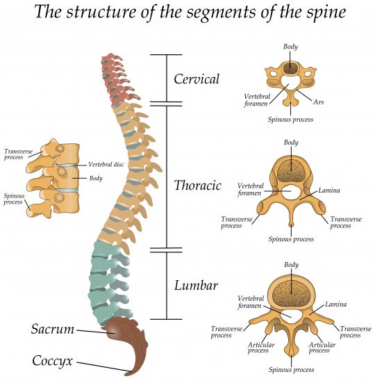
The spinal column is divided into five regions, each with unique characteristics and functions:
- Cervical Spine (C1-C7)
- Location: Neck region.
- Function: Supports the head and allows flexible movement.
- Key Vertebrae:
- C1 (Atlas): Supports the skull.
- C2 (Axis): Enables head rotation.
- Associated Areas: Diaphragm, shoulders, arms, esophagus, and chest.
- Thoracic Spine (T1-T12)
- Location: Mid-back.
- Function: Anchors the rib cage, providing stability.
- Associated Areas: Arms, esophagus, trachea, heart, lungs, liver, gallbladder, and small intestine.
- Lumbar Spine (L1-L5)
- Location: Lower back.
- Function: Bears body weight and supports movement.
- Associated Areas: Legs and feet.
- Sacrum (S1-S5)
- Location: Below the lumbar spine, between the iliac bones.
- Function: Forms the back of the pelvis, connecting to the sacroiliac joints.
- Associated Areas: Bowel, bladder, and sexual function.
- Coccyx (Tailbone)
- Location: Below the sacrum.
- Function: Supports weight when sitting.
- Structure: 3–5 fused bones, also called coccygeal vertebrae.
The vertebral column forms a natural double-S curve when viewed from the side, with inward curves in the cervical and lumbar regions and an outward curve in the thoracic region. This curvature enhances strength, flexibility, and shock absorption.
Sacrum and Coccyx – The Base of the Spine
The sacrum is a triangular bone located below the lumbar spine, connecting to the pelvis via the sacroiliac joints (SI joints). Below the sacrum lies the coccyx, or tailbone, made up of 3–5 fused vertebrae. Though small, the coccyx plays a crucial role in weight distribution while sitting.
Supporting Structures of the Spine
The spinal column is more than just bones. It includes:
- Intervertebral Discs: Fibrocartilage cushions between vertebrae (C3–L5), acting as shock absorbers
- Facet Joints (Zygapophyseal Joints): Paired joints that allow controlled spinal motion
- Spinal Ligaments: Tough bands that stabilize the spine during movement
- Spinal Muscles and Tendons: Provide strength, posture, and movement control
These components work together to maintain spinal alignment and flexibility.
The Connection Between Spinal Nerves and Body Function
Think of the spinal canal as an interstate highway and the foramen as exit ramps. Each set of spinal nerves exits through specific foramina to serve particular areas of the body, enabling both motor function (movement) and sensation (feeling). For instance:
- Cervical Spine Nerves: Associated with the diaphragm (breathing), shoulders, parts of the arms, esophagus, and parts of the chest.
- Thoracic Spine Nerves: Connect to parts of the arm, esophagus, trachea, heart, lungs, liver, gallbladder, and small intestine.
- Lumbar Spine Nerves: Primarily control the legs and feet.
- Sacral Nerves: Responsible for bowel, bladder, and sexual function
The spinal column is the bony structure made up of vertebrae that provides support and protects the spinal cord, which is a delicate bundle of nerves running through the spinal canal. The spinal column is part of the skeletal system, while the spinal cord is part of the central nervous system.
Intervertebral discs act as shock absorbers between the vertebrae and help maintain flexibility and movement in the spine. Healthy discs prevent vertebrae from rubbing against each other and reduce the risk of spinal disorders such as herniated discs or degenerative disc disease.
Yes, spinal misalignment or nerve compression in specific regions of the vertebral column can disrupt signals to internal organs. For example, issues in the thoracic spine may affect lung or heart function, while problems in the sacral spine may impact the bladder or bowel.
Spine specialists use a combination of physical exams, neurological tests, imaging (like MRI or CT scans), and patient history to pinpoint the affected area of the spinal column. Conditions such as sciatica, spinal stenosis, or pinched nerves are identified based on symptoms and diagnostic findings.
Mild spinal degeneration, such as thinning intervertebral discs or minor joint changes, is common with aging. However, advanced degeneration causing pain, nerve compression, or mobility issues may require intervention from a spine doctor or spine surgeon.